| |
The nucleus is the brain of eukaryotic cells.
It is only present in eukaryotic cells (which are eukaryotic
because they have a nucleus) and there is only one of these
organelles in each cell. Usually the nucleus is round and
is the largest organelle in the cell. It is surrounded by
a membrane, called the nuclear envelope, which is similar
to the cell
membrane that encloses the entire cell. The envelope is
riddled with holes, called nuclear pores, that allow specific
materials to pass in and out of the nucleus, just like proteins
in the cell membrane regulate the movement of molecules in
and out of the cell itself. Attached to the nuclear envelope
is the endoplasmic reticulum. The nucleus is surrounded by
the cytoplasm
inside a cell.
|

The nucleus is the pink half-sphere in the center of the cell.
Source: Audesirk, Gerald, and Audesirk, Teresa. Biology:
Life on Earth. |

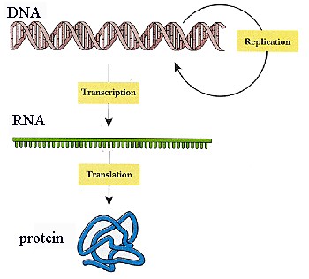
DNA to protein
|
The nucleus houses the DNA (deoxyribonucleic
acid) which stores genetic information for a cell. The DNA
contains instructions for the production of the cell's proteins
and for reproduction. To construct proteins, the DNA is copied
to messenger RNA (ribonucleic acid) in the process called
transcription. The mRNA goes to the ribosomes,
either in the nucleus or in the endoplasmic
reticulum, where the actual construction of the proteins
takes place.
Structurally, the nucleus is composed of three
main parts, the nucleolus, the nuclear envelope, and the chromatin.
The nucleolus contains ribosomes, RNA, DNA, and proteins.
The nucleolus has some of the ribosomes that synthesize proteins
(others are in the endoplasmic reticulum). The chromatin (meaning
"colored substance") contains DNA and proteins formed into
packets of code called chromosomes. When the cell divides,
the chromosomes fold up on themselves, getting wider. The
nuclear envelope is important because it allows the nucleus
to control the rest of the cell, such as by sending out ATP.
The envelope will let molecules like ATP through but will
keep other things in or out, so the nucleus is isolated from
the cytoplasm. |
Endoplasmic Reticulum
Role of the Ribosome
The route from the DNA code to the protein.
 Before cell division, the DNA in our chromosomes replicates
so each daughter cell has an identical set of chromosome.
In addition, the DNA is responsible for coding for all proteins.
Each amino acid is designated by one or more set of triplet
nucleotides. The code is produced from one strand of the DNA
by a process called "transcription". This produces mRNA which
then is sent out of the nucleus where the message is translated
into proteins. This can be done in the cytoplasm on
clusters of ribosomes, called "polyribosomes". Or it
can be done on the membranes of the rough endoplasmic reticulum.
The cartoon to the left shows the basic sequence of transcription
and translational events.
Before cell division, the DNA in our chromosomes replicates
so each daughter cell has an identical set of chromosome.
In addition, the DNA is responsible for coding for all proteins.
Each amino acid is designated by one or more set of triplet
nucleotides. The code is produced from one strand of the DNA
by a process called "transcription". This produces mRNA which
then is sent out of the nucleus where the message is translated
into proteins. This can be done in the cytoplasm on
clusters of ribosomes, called "polyribosomes". Or it
can be done on the membranes of the rough endoplasmic reticulum.
The cartoon to the left shows the basic sequence of transcription
and translational events. |
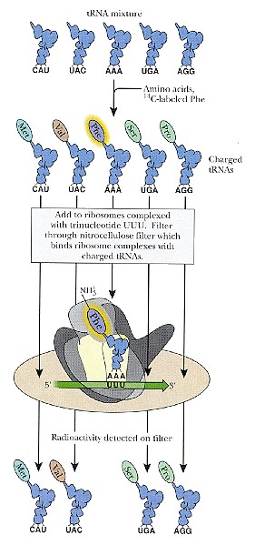
What happens at the site of the ribosome?
 The
code is actually translated on structures that are also made
in the nucleus, called Ribosomes. These ribosomes provide
the structural site where the mRNA sits. The amino acids
for the proteins are carried to the site by "transfer RNAs,".
In the cartoon to the left, these are shown as blue molecules.
Each transfer RNA (tRNA) has a nucleotide triplet which binds
to the complementary sequence on the mRNA (see the three letters
at the bottom of each molecule). The
code is actually translated on structures that are also made
in the nucleus, called Ribosomes. These ribosomes provide
the structural site where the mRNA sits. The amino acids
for the proteins are carried to the site by "transfer RNAs,".
In the cartoon to the left, these are shown as blue molecules.
Each transfer RNA (tRNA) has a nucleotide triplet which binds
to the complementary sequence on the mRNA (see the three letters
at the bottom of each molecule).
The tRNA carries the amino acid at its opposite end. One
can trace and detect binding of a particular tRNA-amino
acid complex to the mRNA by labeling that amino acid.
It will bind to its tRNA. In the case to the left,
Phenylalanine is bound to the tRNA which carries the complementary
base code AAA (adenine-adenine-adenine). This triplet
code would bind to the complementary sequence on mRNA UUU
(uracil X3). The mRNA is shown as a green arrow.
This cartoon shows the selective binding site on the mRNA
which is attached in the ribosome. It also shows the
tRNA carrying the Phenylalanine bound at the site
In this particular assay which uses a polyuracil mRNA, only
phenylalanine-bearing tRNA is bound and detected on the
filter.
Initiation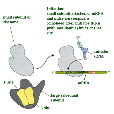
The cartoon shows the initiation of this process. It begins
with the small subunit of the ribosome bound to the mRNA.
An initiator tRNA is attracted to the region (carrying a
methionine. It binds to the triplet code AUG.
This then attracts the large ribosomal subunit which will
bind to the small subunit. Note that it has an A site and
a P site. These are different binding sites for the
tRNAs. The cartoon below describes the next phase
in the process.
Elongation
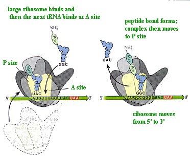 In this cartoon, note that the initiator tRNA complex has
moved to the P site. This leaves the A site open for
the next tRNA. In this case, we have Proline, which
carries the complementary code GGC. Note that its binding
site on the mRNA is CCG. After binding to the A site,
the peptide bond between the methionine and proline forms.
The empty tRNA carrying the MET leaves and the tRNA carrying
the Proline moves to the P site. The ribosome moves to the
next triplet code from 5' to the 3' direction (note arrow
on mRNA). The tRNAs are moving from 3' to the 5' direction
as the ribosome reads the code
In this cartoon, note that the initiator tRNA complex has
moved to the P site. This leaves the A site open for
the next tRNA. In this case, we have Proline, which
carries the complementary code GGC. Note that its binding
site on the mRNA is CCG. After binding to the A site,
the peptide bond between the methionine and proline forms.
The empty tRNA carrying the MET leaves and the tRNA carrying
the Proline moves to the P site. The ribosome moves to the
next triplet code from 5' to the 3' direction (note arrow
on mRNA). The tRNAs are moving from 3' to the 5' direction
as the ribosome reads the code
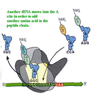 The ribosome continues to read the code from the 5' to the
3' and amino acids are added to the growing peptide chain.
This one shows the tRNA carrying the glycine amino acid
coded by CCA. Its complementary bases are GGU.
The ribosome continues to read the code from the 5' to the
3' and amino acids are added to the growing peptide chain.
This one shows the tRNA carrying the glycine amino acid
coded by CCA. Its complementary bases are GGU.
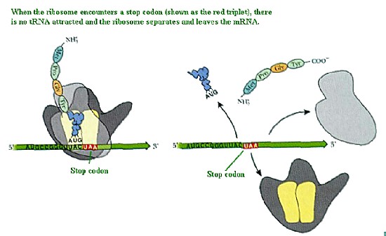
This continues until the stop codon is reached. This
is highlighted in red in this figure and the next figure.
The following cartoon shows what happens when the stop
codon is reached.
End of translation
Clusters of ribosomes may sit on a mRNA and make proteins,
each making a strand of polypeptides. These clusters
are called polyribosomes. When they are free in the cytoplasm,
they are called free polyribosomes (linked by the mRNA).
Or, they may bind to rough endoplasmic reticulum.
Ribosomes are visualized as small (20 X 30 nm) ribonucleoprotein
particles. They are formed from two subunits. As you learned
in the lecture on the nucleolus
, the subunits are produced in the nucleolus in organizing
centers on certain chromosomes. The two ribosomal subunits
leave the nucleus separately through the nuclear
pores . The pores are structured to allow transit of
only the subunits. Whole ribosomes are formed outside in
the cytoplasm. This prevents protein synthesis from occurring
in the nucleus. Why might this be important?
The above photograph shows a group of ribosomes in action.
They are connected by a strand of messenger RNA which runs
between the large and small subunits. They read the 3 nucleotide
code for an amino acid and the appropriate transfer RNA
brings the amino acid to the growing polypeptide chain.
In this photograph, we see the growing peptide chain radiating
at right angles to the mRNA. It extends from the base of
the large ribosomal subunit.
Return
to Menu |
 The
left hand view of this cartoon shows the free polyribosomes
connected by the mRNA. They are arranged in rosettes and these
can be seen in the cytoplasm in conventional electron micrographs.
The right hand view shows the arrangement of polyribosomes
on the rough endoplasmic reticulum. Note that the growing
polypeptide chain (which projects down from the large subunit)
is inserted through the membrane and into the cisterna of
the rough endoplasmic reticulum. The
left hand view of this cartoon shows the free polyribosomes
connected by the mRNA. They are arranged in rosettes and these
can be seen in the cytoplasm in conventional electron micrographs.
The right hand view shows the arrangement of polyribosomes
on the rough endoplasmic reticulum. Note that the growing
polypeptide chain (which projects down from the large subunit)
is inserted through the membrane and into the cisterna of
the rough endoplasmic reticulum.
Return
to Menu

This cartoon shows the binding site on the rough endoplasmic
reticulum. The membrane of the rough endoplasmic has a receptor
that binds the larger subunit of the ribosome. Next to the
receptor is a pore that allows newly synthesized proteins
to enter and be stored initially in the rough endoplasmic
reticulum cisterna or lumen. Note that the ribosomes are
still connected to one another outside the rough endoplasmic
reticulum by the mRNA which runs between the large and small
subunits.
|
 This
electron micrograph shows a high magnification of a longitudinal
section through the rough endoplasmic reticulum. The electron
dense ribosomes are on its outside surface. Inside the sac
(cisterna) is flocculent material, the newly synthesized proteins.
The details of ribosomal structure cannot be appreciated in
this micrograph. They look like small irregular balls on the
outside of the membrane. Note that the sacs of rough endoplasmic
reticulum are bridged by a junction. This is shown diagrammatically
in the following figure. This
electron micrograph shows a high magnification of a longitudinal
section through the rough endoplasmic reticulum. The electron
dense ribosomes are on its outside surface. Inside the sac
(cisterna) is flocculent material, the newly synthesized proteins.
The details of ribosomal structure cannot be appreciated in
this micrograph. They look like small irregular balls on the
outside of the membrane. Note that the sacs of rough endoplasmic
reticulum are bridged by a junction. This is shown diagrammatically
in the following figure. |

The cartoon in this figure shows the rough endoplasmic reticulum
with a bridge adjoining two sacs. In this way, the sacs
communicate and proteins fill the spaces all over the cell.
They even communicate with the inside of the nuclear
envelope. Recall that the outside membrane of the nuclear
envelope is studded with ribosomes and is part of the rough
endoplasmic reticulum. An immunocytochemical labeling protocol,
such as that found in the above
figure, will delineate the reticulum filled with the
newly synthesized proteins.
|
The lysosomes have
sometimes been likened to "The Police Force of the Cell".
Even in a place as small as a cell, we need someone to keep things
in order. But unlike the police, these lysosomes literally eat things
which disturb the natural order of the cell. Lysosomes pick up foreign
invaders such as bacteria, food and old organelles and break them
into small pieces that can hopefully be used again. If they pick
up a really harmful invader, they will eat it up and expel what
is left of it out of the cell so that the debris can be removed
from the body.
The lysosome is able to do this because it is filled with enzymes.
These enzymes are specially made for the lysosome by the rough endoplasmic
reticulum and work only at low pH (highly acidic) levels. The reason
for this is that the enzymes are so strong that they could eat the
whole cell if the lysosome ever let them out. However because they
can only work at low pH levels and the rest of the cell has a neutral
pH level, they can be neutralized if they accidentally escape from
the lysosome.
|
|